Annexin A5
Protein-coding gene in the species Homo sapiens
| ANXA5 | |||||||||||||||||||||||||||||||||||||||||||||||||||
|---|---|---|---|---|---|---|---|---|---|---|---|---|---|---|---|---|---|---|---|---|---|---|---|---|---|---|---|---|---|---|---|---|---|---|---|---|---|---|---|---|---|---|---|---|---|---|---|---|---|---|---|
 | |||||||||||||||||||||||||||||||||||||||||||||||||||
| Identifiers | |||||||||||||||||||||||||||||||||||||||||||||||||||
| Aliases | ANXA5, ANX5, ENX2, HEL-S-7, PP4, RPRGL3, annexin A5, CPB-I, VAC-alph | ||||||||||||||||||||||||||||||||||||||||||||||||||
| External IDs | OMIM: 131230; MGI: 106008; HomoloGene: 20312; GeneCards: ANXA5; OMA:ANXA5 - orthologs | ||||||||||||||||||||||||||||||||||||||||||||||||||
| |||||||||||||||||||||||||||||||||||||||||||||||||||
| |||||||||||||||||||||||||||||||||||||||||||||||||||
| |||||||||||||||||||||||||||||||||||||||||||||||||||
| |||||||||||||||||||||||||||||||||||||||||||||||||||
| |||||||||||||||||||||||||||||||||||||||||||||||||||
| Wikidata | |||||||||||||||||||||||||||||||||||||||||||||||||||
| |||||||||||||||||||||||||||||||||||||||||||||||||||


Annexin A5 (or annexin V) is a cellular protein in the annexin group. In flow cytometry, annexin V is commonly used to detect apoptotic cells by its ability to bind to phosphatidylserine, a marker of apoptosis when it is on the outer leaflet of the plasma membrane. The function of the protein is unknown; however, annexin A5 has been proposed to play a role in the inhibition of blood coagulation by competing for phosphatidylserine binding sites with prothrombin and also to inhibit the activity of phospholipase A1. These properties have been found by in vitro experiments.
Pathology
Antibodies directed against annexin A5 are found in patients with a disease called the antiphospholipid syndrome (APS), a thrombophilic disease associated with autoantibodies against phospholipid compounds.
Annexin A5 forms a shield around negatively charged phospholipid molecules. The formation of an annexin A5 shield blocks the entry of phospholipids into coagulation (clotting) reactions. In the antiphospholipid antibody syndrome, the formation of the shield is disrupted by antibodies. Without the shield, there is an increased quantity of phospholipid molecules on cell membranes, speeding up coagulation reactions and causing the blood-clotting characteristic of the antiphospholipid antibody syndrome.
Annexin A5 showed upregulation in papillary thyroid carcinoma.[5]
Laboratory use
Annexin A5 is used as a non-quantitative probe to detect cells that have expressed phosphatidylserine (PS) on the cell surface, an event found in apoptosis as well as other forms of cell death.[6][7][8] Platelets also expose PS and PE on their surface when activated, which serves as binding site for various coagulation factors.
The annexin A5 affinity assay typically uses a conjugate of annexin V and a fluorescent or enzymatic label, biotin or other tags, or a radioelement, in a suitable buffer (annexin V binding to aminophospholipids is Ca2+ dependent). The assay combines annexin V staining of PS and PE membrane events with the staining of DNA in the cell nucleus with propidium iodide (PI) or 7-Aminoactinomycin D (AAD-7), distinguishing viable cells from apoptotic cells and necrotic cells.[9] Detection occurs by flow cytometry or a fluorescence microscope.
Interactions
Annexin A5 has been shown to interact with Kinase insert domain receptor[10] and Integrin, beta 5.[11]
References
- ^ a b c GRCh38: Ensembl release 89: ENSG00000164111 – Ensembl, May 2017
- ^ a b c GRCm38: Ensembl release 89: ENSMUSG00000027712 – Ensembl, May 2017
- ^ "Human PubMed Reference:". National Center for Biotechnology Information, U.S. National Library of Medicine.
- ^ "Mouse PubMed Reference:". National Center for Biotechnology Information, U.S. National Library of Medicine.
- ^ Sofiadis A, Becker S, Hellman U, Hultin-Rosenberg L, Dinets A, Hulchiy M, Zedenius J, Wallin G, Foukakis T, Höög A, Auer G, Lehtiö J, Larsson C (Apr 2012). "Proteomic profiling of follicular and papillary thyroid tumors". European Journal of Endocrinology. 166 (4): 657–67. doi:10.1530/EJE-11-0856. PMC 3315832. PMID 22275472.
- ^ Meers P and Mealy T (1994). "Phospholipid determinants for annexin V binding sites and the role of tryptophan". Biochemistry. 33 (19): 5829–37. doi:10.1021/bi00185a022. PMID 8180211.
- ^ Koopman G, Reutelingsperger CP, Kuijten GA, Keehnen RM, Pals ST, van Oers MH (Sep 1994). "Annexin V for flow cytometric detection of phosphatidylserine expression on B cells undergoing apoptosis". Blood. 84 (5): 1415–20. doi:10.1182/blood.V84.5.1415.1415. PMID 8068938.
- ^ Vermes I, Haanen C, Steffens-Nakken H, Reutelingsperger C (Jul 1995). "A novel assay for apoptosis. Flow cytometric detection of phosphatidylserine expression on early apoptotic cells using fluorescein labelled Annexin V". Journal of Immunological Methods. 184 (1): 39–51. doi:10.1016/0022-1759(95)00072-I. PMID 7622868.
- ^ Annexin-FP488 fluorescent staining protocol at Interchim
- ^ Wen Y, Edelman JL, Kang T, Sachs G (May 1999). "Lipocortin V may function as a signaling protein for vascular endothelial growth factor receptor-2/Flk-1". Biochemical and Biophysical Research Communications. 258 (3): 713–21. doi:10.1006/bbrc.1999.0678. PMID 10329451.
- ^ Cardó-Vila M, Arap W, Pasqualini R (May 2003). "Alpha v beta 5 integrin-dependent programmed cell death triggered by a peptide mimic of annexin V". Molecular Cell. 11 (5): 1151–62. doi:10.1016/S1097-2765(03)00138-2. PMID 12769841.
Further reading
- Cederholm A, Frostegård J (Jun 2007). "Annexin A5 as a novel player in prevention of atherothrombosis in SLE and in the general population". Annals of the New York Academy of Sciences. 1108 (1): 96–103. Bibcode:2007NYASA1108...96C. doi:10.1196/annals.1422.011. PMID 17893975. S2CID 29296971.
- Schlaepfer DD, Jones J, Haigler HT (Feb 1992). "Inhibition of protein kinase C by annexin V". Biochemistry. 31 (6): 1886–91. doi:10.1021/bi00121a043. PMID 1310621.
- Huber R, Berendes R, Burger A, Schneider M, Karshikov A, Luecke H, Römisch J, Paques E (Feb 1992). "Crystal and molecular structure of human annexin V after refinement. Implications for structure, membrane binding and ion channel formation of the annexin family of proteins". Journal of Molecular Biology. 223 (3): 683–704. doi:10.1016/0022-2836(92)90984-R. PMID 1311770.
- Kirsch T, Pfäffle M (Sep 1992). "Selective binding of anchorin CII (annexin V) to type II and X collagen and to chondrocalcin (C-propeptide of type II collagen). Implications for anchoring function between matrix vesicles and matrix proteins". FEBS Letters. 310 (2): 143–7. doi:10.1016/0014-5793(92)81316-E. PMID 1397263. S2CID 9498732.
- Dawson SJ, White LA (May 1992). "Treatment of Haemophilus aphrophilus endocarditis with ciprofloxacin". The Journal of Infection. 24 (3): 317–20. doi:10.1016/S0163-4453(05)80037-4. PMID 1602151.
- Tait JF, Frankenberry DA, Shiang R, Murray JC, Adler DA, Disteche CM (1992). "Chromosomal localization of the human gene for annexin V (placental anticoagulant protein I) to 4q26----q28". Cytogenetics and Cell Genetics. 57 (4): 187–92. doi:10.1159/000133143. PMID 1683830.
- Huber R, Römisch J, Paques EP (Dec 1990). "The crystal and molecular structure of human annexin V, an anticoagulant protein that binds to calcium and membranes". The EMBO Journal. 9 (12): 3867–74. doi:10.1002/j.1460-2075.1990.tb07605.x. PMC 552154. PMID 2147412.
- Huber R, Schneider M, Mayr I, Römisch J, Paques EP (Nov 1990). "The calcium binding sites in human annexin V by crystal structure analysis at 2.0 A resolution. Implications for membrane binding and calcium channel activity". FEBS Letters. 275 (1–2): 15–21. doi:10.1016/0014-5793(90)81428-Q. PMID 2148156. S2CID 8975064.
- Maurer-Fogy I, Reutelingsperger CP, Pieters J, Bodo G, Stratowa C, Hauptmann R (Jul 1988). "Cloning and expression of cDNA for human vascular anticoagulant, a Ca2+-dependent phospholipid-binding protein". European Journal of Biochemistry. 174 (4): 585–92. doi:10.1111/j.1432-1033.1988.tb14139.x. PMID 2455636.
- Rothhut B, Coméra C, Cortial S, Haumont PY, Diep Le KH, Cavadore JC, Conard J, Russo-Marie F, Lederer F (Nov 1989). "A 32 kDa lipocortin from human mononuclear cells appears to be identical with the placental inhibitor of blood coagulation". The Biochemical Journal. 263 (3): 929–35. doi:10.1042/bj2630929. PMC 1133519. PMID 2532007.
- Schlaepfer DD, Mehlman T, Burgess WH, Haigler HT (Sep 1987). "Structural and functional characterization of endonexin II, a calcium- and phospholipid-binding protein". Proceedings of the National Academy of Sciences of the United States of America. 84 (17): 6078–82. Bibcode:1987PNAS...84.6078S. doi:10.1073/pnas.84.17.6078. PMC 299011. PMID 2957692.
- Funakoshi T, Heimark RL, Hendrickson LE, McMullen BA, Fujikawa K (Aug 1987). "Human placental anticoagulant protein: isolation and characterization". Biochemistry. 26 (17): 5572–8. doi:10.1021/bi00391a053. PMID 2960376.
- Iwasaki A, Suda M, Nakao H, Nagoya T, Saino Y, Arai K, Mizoguchi T, Sato F, Yoshizaki H, Hirata M (Nov 1987). "Structure and expression of cDNA for an inhibitor of blood coagulation isolated from human placenta: a new lipocortin-like protein". Journal of Biochemistry. 102 (5): 1261–73. doi:10.1093/oxfordjournals.jbchem.a122165. PMID 2963810.
- Funakoshi T, Hendrickson LE, McMullen BA, Fujikawa K (Dec 1987). "Primary structure of human placental anticoagulant protein". Biochemistry. 26 (25): 8087–92. doi:10.1021/bi00399a011. PMID 2964863.
- Kaplan R, Jaye M, Burgess WH, Schlaepfer DD, Haigler HT (Jun 1988). "Cloning and expression of cDNA for human endonexin II, a Ca2+ and phospholipid binding protein". The Journal of Biological Chemistry. 263 (17): 8037–43. doi:10.1016/S0021-9258(18)68438-8. PMID 2967291.
- Grundmann U, Abel KJ, Bohn H, Löbermann H, Lottspeich F, Küpper H (Jun 1988). "Characterization of cDNA encoding human placental anticoagulant protein (PP4): homology with the lipocortin family". Proceedings of the National Academy of Sciences of the United States of America. 85 (11): 3708–12. Bibcode:1988PNAS...85.3708G. doi:10.1073/pnas.85.11.3708. PMC 280287. PMID 2967495.
- Pepinsky RB, Tizard R, Mattaliano RJ, Sinclair LK, Miller GT, Browning JL, Chow EP, Burne C, Huang KS, Pratt D (Aug 1988). "Five distinct calcium and phospholipid binding proteins share homology with lipocortin I". The Journal of Biological Chemistry. 263 (22): 10799–811. doi:10.1016/S0021-9258(18)38041-4. PMID 2968983.
- Ahn NG, Teller DC, Bienkowski MJ, McMullen BA, Lipkin EW, de Haën C (Dec 1988). "Sedimentation equilibrium analysis of five lipocortin-related phospholipase A2 inhibitors from human placenta. Evidence against a mechanistically relevant association between enzyme and inhibitor". The Journal of Biological Chemistry. 263 (35): 18657–63. doi:10.1016/S0021-9258(18)37335-6. PMID 2974032.
- Demange P, Voges D, Benz J, Liemann S, Göttig P, Berendes R, Burger A, Huber R (Jul 1994). "Annexin V: the key to understanding ion selectivity and voltage regulation?". Trends in Biochemical Sciences. 19 (7): 272–6. doi:10.1016/0968-0004(94)90002-7. PMID 7519374.
- Fernández MP, Morgan RO, Fernández MR, Carcedo MT (Nov 1994). "The gene encoding human annexin V has a TATA-less promoter with a high G+C content". Gene. 149 (2): 253–60. doi:10.1016/0378-1119(94)90157-0. PMID 7958998.
External links
- Annexin+A5 at the U.S. National Library of Medicine Medical Subject Headings (MeSH)
- Human ANXA5 genome location and ANXA5 gene details page in the UCSC Genome Browser.
- v
- t
- e
PDB gallery
-
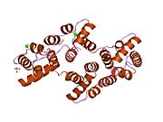
 1a8a: RAT ANNEXIN V COMPLEXED WITH GLYCEROPHOSPHOSERINE
 -
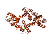
 1a8b: RAT ANNEXIN V COMPLEXED WITH GLYCEROPHOSPHOETHANOLAMINE
 -
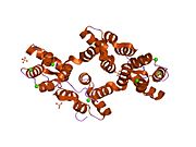
 1anw: THE EFFECT OF METAL BINDING ON THE STRUCTURE OF ANNEXIN V AND IMPLICATIONS FOR MEMBRANE BINDING
 -
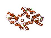
 1anx: THE CRYSTAL STRUCTURE OF A NEW HIGH-CALCIUM FORM OF ANNEXIN V
 -
 1avh: CRYSTAL AND MOLECULAR STRUCTURE OF HUMAN ANNEXIN V AFTER REFINEMENT. IMPLICATIONS FOR STRUCTURE, MEMBRANE BINDING AND ION CHANNEL FORMATION OF THE ANNEXIN FAMILY OF PROTEINS
 -
 1avr: CRYSTAL AND MOLECULAR STRUCTURE OF HUMAN ANNEXIN V AFTER REFINEMENT. IMPLICATIONS FOR STRUCTURE, MEMBRANE BINDING AND ION CHANNEL FORMATION OF THE ANNEXIN FAMILY OF PROTEINS
 -
 1bc0: RECOMBINANT RAT ANNEXIN V, W185A MUTANT
 -
 1bc1: RECOMBINANT RAT ANNEXIN V, QUADRUPLE MUTANT (T72K, S144K, S228K, S303K)
 -
 1bc3: RECOMBINANT RAT ANNEXIN V, TRIPLE MUTANT (T72K, S144K, S228K)
 -
 1bcw: RECOMBINANT RAT ANNEXIN V, T72A MUTANT
 -
 1bcy: RECOMBINANT RAT ANNEXIN V, T72K MUTANT
 -
 1bcz: RECOMBINANT RAT ANNEXIN V, T72S MUTANT
 -
 1g5n: ANNEXIN V COMPLEX WITH HEPARIN OLIGOSACCHARIDES
 -
 1hak: CRYSTAL STRUCTURE OF RECOMBINANT HUMAN PLACENTAL ANNEXIN V COMPLEXED WITH K-201 AS A CALCIUM CHANNEL ACTIVITY INHIBITOR
 -
 1hvd: STRUCTURAL AND ELECTROPHYSIOLOGICAL ANALYSIS OF ANNEXIN V MUTANTS. MUTAGENESIS OF HUMAN ANNEXIN V, AN IN VITRO VOLTAGE-GATED CALCIUM CHANNEL, PROVIDES INFORMATION ABOUT THE STRUCTURAL FEATURES OF THE ION PATHWAY, THE VOLTAGE SENSOR AND THE ION SELECTIVITY FILTER
 -
 1hve: STRUCTURAL AND ELECTROPHYSIOLOGICAL ANALYSIS OF ANNEXIN V MUTANTS. MUTAGENESIS OF HUMAN ANNEXIN V, AN IN VITRO VOLTAGE-GATED CALCIUM CHANNEL, PROVIDES INFORMATION ABOUT THE STRUCTURAL FEATURES OF THE ION PATHWAY, THE VOLTAGE SENSOR AND THE ION SELECTIVITY FILTER
 -
 1hvf: STRUCTURAL AND ELECTROPHYSIOLOGICAL ANALYSIS OF ANNEXIN V MUTANTS. MUTAGENESIS OF HUMAN ANNEXIN V, AN IN VITRO VOLTAGE-GATED CALCIUM CHANNEL, PROVIDES INFORMATION ABOUT THE STRUCTURAL FEATURES OF THE ION PATHWAY, THE VOLTAGE SENSOR AND THE ION SELECTIVITY FILTER
 -
 1hvg: STRUCTURAL AND ELECTROPHYSIOLOGICAL ANALYSIS OF ANNEXIN V MUTANTS. MUTAGENESIS OF HUMAN ANNEXIN V, AN IN VITRO VOLTAGE-GATED CALCIUM CHANNEL, PROVIDES INFORMATION ABOUT THE STRUCTURAL FEATURES OF THE ION PATHWAY, THE VOLTAGE SENSOR AND THE ION SELECTIVITY FILTER
 -
 1n41: Crystal Structure of Annexin V K27E Mutant
 -
 1n42: Crystal Structure of Annexin V R149E Mutant
 -
 1n44: Crystal Structure of Annexin V R23E Mutant
 -
 1sav: HUMAN ANNEXIN V WITH PROLINE SUBSTITUTION BY THIOPROLINE
 -
 2ie6: Annexin V under 2.0 MPa pressure of xenon
 -
 2ie7: Annexin V under 2.0 MPa pressure of nitrous oxide
 -
 2ran: RAT ANNEXIN V CRYSTAL STRUCTURE: CA2+-INDUCED CONFORMATIONAL CHANGES


