Supraorbital foramen
Opening above the eye socket, below the forehead
| Supraorbital foramen | |
|---|---|
 The skull from the front. ("Supraorbital foramen" is top caption for right side white-box.) | |
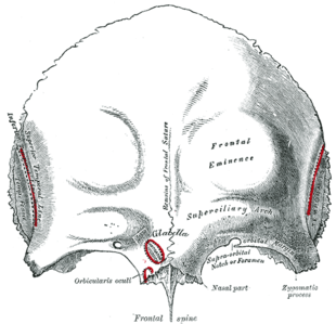 Frontal bone. Outer surface. ("Supraorbital notch or foramen" labeled at lower right arch.) | |
| Details | |
| Identifiers | |
| Latin | foramen supraorbitale |
| TA98 | A02.1.03.009 |
| TA2 | 527 |
| FMA | 57412 |
| Anatomical terms of bone [edit on Wikidata] | |
The supraorbital foramen, is a bony elongated opening located above the orbit (eye socket) and under the forehead. It is part of the frontal bone of the skull. The supraorbital foramen lies directly under the eyebrow. In some people this foramen is incomplete and is then known as the supraorbital notch.[1]
Structure
The supraorbital foramen is a small groove at superior and medial margin of the orbit in the frontal bone. It is part of the frontal bone of the skull.[2] It arches transversely below the superciliary arches and is the upper part of the brow ridge. It is thin and prominent in its lateral two-thirds, but rounded in its medial third.[3] Between these two parts, the supraorbital nerve, the supraorbital artery, and the supraorbital vein pass. The supraorbital nerve divides into superficial and deep branches after it has left the supraorbital foramen.[4]
Additional images
-
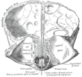 Frontal bone. Inner surface.
 -
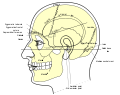 Side view of head, showing surface relations of bones.
 -
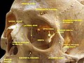 Cranium. Supraorbital foramen.


See also
- Foramina of skull
- Frontal bone
- Supraorbital ridge
References
![]() This article incorporates text in the public domain from page 186 of the 20th edition of Gray's Anatomy (1918)
This article incorporates text in the public domain from page 186 of the 20th edition of Gray's Anatomy (1918)
- ^ Tortora, G; Derrickson, B (2011). Principles of anatomy & physiology (13th. ed.). Wiley. p. 214. ISBN 9780470646083.
- ^ Irby, Nita (2014). "23 - Ophthalmology". Equine Emergencies (4th ed.). Saunders. pp. 379–417. doi:10.1016/B978-1-4557-0892-5.00023-4. ISBN 978-1-4557-0892-5.
- ^ Forehead Anatomy at eMedicine
- ^ Knize, David M. (1995). "A study of the supraorbital nerve". Plastic and Reconstructive Surgery. 96 (3): 564–9. doi:10.1097/00006534-199509000-00007. PMID 7638280.
External links
- "Anatomy diagram: 34256.000-1". Roche Lexicon - illustrated navigator. Elsevier. Archived from the original on 2012-12-27.
- v
- t
- e
Neurocranium of the skull
| Squamous part | |
|---|---|
| Lateral parts | |
| Basilar part |
|
| Other |
| Squamous part |
|
|---|---|
| Orbital part |
| Squamous part | |
|---|---|
| Mastoid part | |
| Petrous part |
|
| Tympanic part |
| Surfaces |
|
|---|---|
| Great wings | |
| Small wings | |
| Pterygoid processes | |
| Other |
| Plates | |
|---|---|
| Surfaces |
|
| Labyrinth |
|
Portal:
 Anatomy

| Authority control databases |
|
|---|


